The nematode or roundworm itself (Nematoda) is a protostome, a protocoelomate, a bilaterally symmetrical moult.
spread. Nematodes are one of the most widespread animal types, able to colonize a variety of habitats - from interstitium (the spaces between grains of sand) and moss communities to Arctic ice (e.g.Tristis Melnikovichandred worm, found in the thickness of multi-year ice in the central Arctic Ocean).Researchers are particularly interested in parasitic nematodes because of their diverse hosts.
Building plans. The body is thin, spindle-shaped, tapering toward both ends, and the cross-section is round.The mouth is located at the front end, and the anus (anus) is located at the rear end.The outside of the body is covered with multiple layers of elastic stratum corneum - a non-cellular structure secreted by the subcutaneous tissue.The hypodermis, or epidermis, lies beneath the stratum corneum.The muscle is represented by a layer of longitudinal oblique muscle fibers.The primary body cavity (split body cavity) lacks its own epithelial lining and is filled with fluid.
digestive system. The oral opening at the front end of the body is surrounded by projections - lips (usually three) and leads to a muscular ectodermal pharynx with a triangular cavity.The pharynx leads from a single layer of columnar epithelium to the endodermal midgut.Next is the short ectodermal hindgut, which leads to the anus.
excretory system. The excretory organs are single-cell glands that replace the protonephros.There is usually a cervical gland in the front of the body from which a short excretory duct emanates.There is also the "storage kidney" - a phagocytic organ that accumulates insoluble metabolic products that are not excreted from the body.
Circulatory and respiratory systems. These systems are missing.Breathing occurs through the skin.Anaerobic metabolism is also possible (anaerobic breakdown of glycogen in parasites to butyrate and valerate).
nervous system. The nervous system is of the ladder type.Represented by a neural ring and six longitudinal trunks.Two nerve trunks running along the ventral and dorsal lines are more powerful and connected by a semicircular neural bridge (commissure).
sense organs. Around the mouth are papillae and setae - organs of touch.Some representatives of the ocean possess primitive eyes - pigmented spots.Amphibian chemosensory organs often have the shape of pockets, spirals, or slits.They are located on either side of the head and are especially developed in males as they aid in the search for females.
reproduction and development. Nematodes are dioecious animals.The internal reproductive organs are paired and have a tubular structure.Reproduction is only sexual.Sexual dimorphism is evident: females are larger and males have a curved rear end.Fertilization is internal and viviparous occurs.During development, nematodes undergo four larval stages, separated by molting, accompanied by shedding of the cuticle.Some species (including the famousCaenorhabditis elegans) Under unfavorable conditions, it enters the so-called "dauer" stage - a dormant larvae.
parasitic. Of the more than 24,000 nematode species currently described, approximately half are parasitic.They can affect almost all tissues and organs: connective tissue, muscles, blood and lymphatic vessels, gonads, sensory organs, and body cavities.These include both ectoparasites of plants, vertebrates, and invertebrates, as well as other nematodes and even protozoa.
The following is a description of the most important representatives of roundworms from the perspective of medical parasitology.
human roundworm(roundworms)
Appearance.Both ends of the body are pointed and pinkish white.Dimensions: males - 15-25 cm, females - 20-40 cm.The body is covered with ten flexible layers of cuticle that protect the host from mechanical stress and digestive enzymes.
spread. The species is cosmopolitan - found throughout the world, but different countries have different proportions of people infected.In Japan, for example, more than 90% of the population is infected with roundworms due to the use of human waste as fertilizer.Roundworms are less common in areas with hot, dry climates.
life cycle.Development proceeds without changing owners.Adult worms live in the small intestine and cause ascariasis.A person is typically infected with dozens of roundworms (900 have been recorded).Lifespan in the intestine is about one year.Like other nematodes, roundworms are dioecious.Sexually mature females lay approximately 200,000 oval-shaped eggs per day, which are released into the external environment in their feces.Roundworms are classified as soil worms - they require soil to develop their larval stage.Larvae develop in the eggs when exposed to favorable conditions (moist soil with a temperature of approximately 25°C and sufficient oxygen).The development period ranges from 16 days to several months, depending on temperature.The eggs containing larvae can be considered invasive.
Infection occurs when eggs are ingested in food or water; transmission does not occur directly from person to person.In the intestine, the larvae burrow through the intestinal wall, enter the blood vessels and liver, and then migrate through the inferior vena cava to the right atrium and right ventricle.From the latter, the larvae move through the pulmonary circulation to the lungs, where they move from the blood to the alveoli, bronchi, trachea and oral cavity.Secondary infection occurs in the oral cavity: the larvae are swallowed, enter the intestines, and become sexually mature after three months.The process of "growing up" in nematodes involves shedding their skin (usually four).
Clinical manifestations of ascariasis. During the migratory stage of ascariasis, coughing (which helps the larvae enter the throat), chest pain, allergic reactions, and fever occur.
During the intestinal stage, damage to the intestinal mucosa and poisoning by toxic metabolites occur.Symptoms: Nausea, vomiting, bowel disturbances, loss of appetite.
Long-term effects of infection: general decrease in performance, sleep disturbance.When the worms crawl into the bile ducts and respiratory tract, the results can be fatal.In addition, roundworm larvae can enter the brain (e.g., from the inferior vena cava to the superior vena cava and then along the brachiocephalic vein), causing meningoencephalitis with associated migraines.
prevention. Wash your hands before eating and preparing food.Wash vegetables and fruits.Flies also carry eggs, so using Velcro and the like to combat these dipterans can also help prevent ascariasis.
Interesting facts. Studies have shown that roundworm infection has a positive effect on alleviating the symptoms of autoimmune diseases and improving female fertility.Scientists attribute this to the parasite's effect on the immune system by affecting T-cell levels in the body, but too little is known about the mechanism to draw firm conclusions.
Pinworm(pinworm)
Appearance. Gray-white nematodes, males 2-5 mm long, females 8-14 mm long.The tail end is pointed (hence the name).At the front of the body, the characteristic swelling of the esophagus is evident.
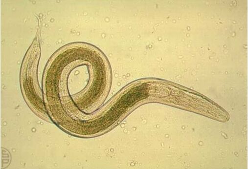
life cycle.Pinworms live in the small intestine and lower part of the large intestine, causing pinworm disease.Lifespan is 1-2 months.The front end of the pinworm is attached to the intestinal wall.Sexually mature females emerge from the large intestine through the anus, lay 5 to 15,000 eggs on the skin near the anus, and then die.
The crawling out of the female is accompanied by itching.When the skin is scratched, the eggs are transferred to the hands etc.Flies are also involved in egg transfer.Infection occurs through ingestion.Larvae hatch from eggs that enter the intestines.
Epidemiology and clinical manifestations of enterobiasis. Pinworms are widespread, especially among children, due to non-compliance with personal hygiene rules and "crowding" in kindergartens and schools.Transmitted from person to person without intermediate host.Reduce the effectiveness of vaccination.
Symptoms: abdominal pain, loss of appetite, headache, allergic symptoms, perianal itching (leading to sleep disturbance and increased irritability).
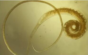
Trichinella spiralis(Trichinella spiralis)
describe.Small nematodes are 2-4 mm long.Parasitic on the mucosa of the small intestine.Distributed in Eurasia and North America.
life cycle. In order for Trichinella spiralis to grow, the host must be changed.Usually these are wild animals (foxes, wolves, bears, wild boars), as well as people and livestock.The female is anchored in the intestinal epithelium by the anterior end of the body and gives birth to 1-2000 larvae.Ovoviviparity is typical: hatching of larvae from eggs occurs in the female reproductive tract.Larvae are transported throughout the body via blood and lymphatic vessels and settle in striated muscles.At this stage, they have a stylet that they use to destroy muscle tissue, causing the host to form a capsule, curled up, in which they will inhabit in the future.After a few months, the capsules are soaked in lime.The muscular trichinella spiralis can persist for years, even after its owner dies and the body decomposes.
Once in the stomach of a new host (after eating the corpse of the previous host), the larvae are released from the capsule, penetrate the mucous membrane, and undergo four molts over several days to transform into adult worms.
Clinical manifestations of trichinellosis. Increased body temperature, facial swelling, muscle pain, allergic reactions.
prevention. Trichinellosis is spread through food through contaminated meat.Therefore, to prevent this disease, meat must be veterinary checked and properly prepared - boiled for 2-3 hours.Cooking methods such as smoking and salting will not kill Trichinella spiralis.
Whipworm(Trichocephalus trichurus)
Appearance.The body of the insect is white and about 4 cm long.The front end is thin, reminiscent of hair (hence the name).
spread.They prefer countries with humid and warm climates.
life cycle.This worm lives in the beginning of the large intestine and is only found in humans.Causes whipworm disease.How many years does a person have?The thin end penetrates the thickness of the intestinal wall mucosa.It feeds on tissue fluid and blood.
The female lays 1-3,000 eggs, which are released into the external environment with the feces.Like roundworms, whipworms are related to soil worms: in order for the eggs to be invasive, they need to remain in the soil at a certain humidity and temperature (25-30°C) for a month.Thereafter, infection occurs when the eggs are swallowed; the larvae emerge from the host's intestines, penetrate the intestinal villi and grow there for about a week.Then, after destroying the villi, they enter the intestinal lumen and reach the large intestine, where they settle and reach maturity within a month.
Clinical manifestations of trichocephaly. The worm damages the colonic mucosa and causes intoxication of the host through waste products.Whipworms are hemophagocytic and therefore can cause anemia.Trichocephaly is associated with abdominal pain, headache, and dizziness.Because whipworms attach to the intestinal wall, they are more difficult to remove from the host than other parasites.
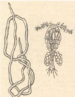
Rishta(guinea worm)
Appearance.A thin white nematode, females are 30-120 cm long and males are no longer than 4 cm.There is a small spine on the tail.
Distribution:Tropical countries in Asia and Africa.
life cycle.Infection can occur when drinking unboiled water that contains copepods.The crustaceans in the stomach die under the influence of hydrochloric acid, but the Guinea worm larvae survive and spread throughout the body through the lymphatic system.They then enter the body cavity, where they molt and reach sexual maturity.After mating, the male dies and the female enters the subcutaneous tissue, forming a purulent abscess accompanied by burning and pain.Cold water provides the best pain relief.
The development of eggs forces the female to start moving the "head" forward towards the surface of the skin, leaving along its path an inflammatory process that turns into a purulent abscess, which then ruptures.When the female's uterus enters the water, it ruptures and the larvae that hatch from the eggs emerge.To ensure that development is not interrupted, the larvae must infect a cyclops crustacean that serves as an intermediate host.Those larvae left in the water will die.After the crustacean is ingested by its final host, under the influence of gastric acid, the crustacean dissolves and the larvae easily enter the intestine, pass through the intestinal wall and eventually enter the lymph nodes, where the developmental cycle continues.The disease caused by dracunculiasis is called dracunculiasis.
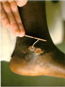
Guinea worm disease.The incubation period lasts up to nine months and ends when the female becomes sexually mature.In people who already have dracunculiasis, this is when purulent abscesses begin to form.The only salvation from the pain is the pond.Relief is immediate, but upon contact with water, the bubbles burst and Guinea worms drop their larvae into the water.The crustaceans eat them and the life cycle begins again.
When treating dracunculiasis, an incision is usually made at the site of the blister and the worms are gradually pulled out and wrapped around a stick.This takes days, sometimes weeks (you have to pull the worm out slowly and carefully so it doesn't burst).Some believe that the appearance of the Guinea worm wrapped around a stick became the prototype for medical symbolism - the staff of Asclepius entwined with a snake.
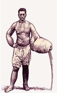
Bancroft silk (filariasis) or Bancroft thread(Utzerella bancrofti)
Appearance.White nematodes, females are 10 cm long and males are 4 cm long.
distribute. Tropical and subtropical areas of Asia, Africa, Central and South America.
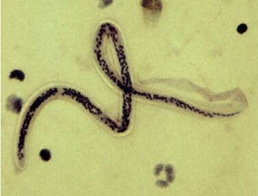
life cycle. Adult worms usually occur in lymph glands and blood vessels, blocking lymphatic drainage and causing persistent swelling.The female produces larvae - nocturnal microfilariae, which appear in the peripheral blood at night and penetrate deeply into the body (entering the pulmonary vessels and kidneys) during the day.This is because the intermediate host is a mosquito, which usually feeds on blood in the evening and at night.The larvae enter the mosquito's stomach and then into the body cavity, where they grow and then gather near the proboscis and are transmitted to humans by sucking blood.Bancroft silk causes elephantiasis, or elephantiasis, or elephantiasis.It is important to note that the disease can also be caused by other nematodes.
Clinical manifestations and treatment of elephantiasis. Swelling in any part of the body is caused by hyperplasia (painful growth) of the skin and subcutaneous tissue, caused by inflammatory thickening of lymphatic walls and stagnation of lymphatic fluid due to blockage of lymphatic vessels by adult Bancroft filamentous individuals.The skin on the affected area of the body is covered with ulcers.
Treatment for elephantiasis aims to improve fluid outflow.Using anthelmintics is effective.In later stages, surgery may be required.